Contents
- 🧲 What Exactly is an MRI Machine?
- 🔬 How Does the Magic Happen? The Physics Explained
- 💡 Key Components: The Anatomy of an MRI Scanner
- 🏥 Who Needs an MRI and Why?
- ⚡ MRI vs. Other Imaging: When to Choose What
- 📈 The Evolution of MRI: From Giant Magnets to AI
- ⚠️ Safety First: What to Know Before Your Scan
- 💰 Cost Considerations and Insurance
- 🌟 Ratings and Reviews: What Patients Experience
- 🚀 The Future of MRI: Faster, Smarter, Smaller?
- Frequently Asked Questions
- Related Topics
Overview
Magnetic Resonance Imaging (MRI) machines are sophisticated diagnostic tools that utilize powerful magnetic fields and radio waves to generate detailed cross-sectional images of the body's internal structures. Unlike X-rays or CT scans, MRI doesn't rely on ionizing radiation, making it a safer option for repeated imaging and for sensitive populations. The core components include a superconducting magnet, radiofrequency coils for transmitting and receiving signals, and a gradient system for spatial encoding. The patient lies within the bore of the magnet, and the interaction of hydrogen protons with the magnetic field and radio pulses creates signals that are processed into images, revealing soft tissue detail with exceptional clarity. Understanding the technology behind MRI is crucial for appreciating its diagnostic power, its limitations, and the ongoing advancements shaping its future in healthcare.
🧲 What Exactly is an MRI Machine?
An Magnetic Resonance Imaging scanner is a sophisticated medical device that uses powerful magnetic fields and radio waves to create detailed cross-sectional images of the body's internal structures. Unlike X-rays or CT scans, it doesn't use ionizing radiation, making it a preferred choice for imaging soft tissues, organs, and the brain. These machines are typically found in hospitals and specialized imaging centers, serving diagnostic purposes across a vast range of medical specialties, from neurology to orthopedics. The core function is to visualize what's happening beneath the surface without invasive procedures, offering crucial insights for diagnosis and treatment planning. Understanding the basics of how these complex machines operate can demystify the process for patients and healthcare professionals alike.
🔬 How Does the Magic Happen? The Physics Explained
The fundamental principle behind MRI lies in nuclear magnetic resonance. Protons within the body's water molecules, primarily hydrogen, act like tiny magnets. When placed in a strong magnetic field generated by the MRI machine, these protons align themselves. Radiofrequency pulses are then emitted, knocking these aligned protons out of equilibrium. As the protons realign with the magnetic field, they release energy that is detected by the scanner's receiver coils. Different tissues have varying amounts of water and protons, and they realign at different rates, producing distinct signals. These signals are then processed by a powerful computer to construct detailed anatomical images, a process that has been refined since the initial discoveries by Felix Bloch and Edward Purcell in the 1940s.
💡 Key Components: The Anatomy of an MRI Scanner
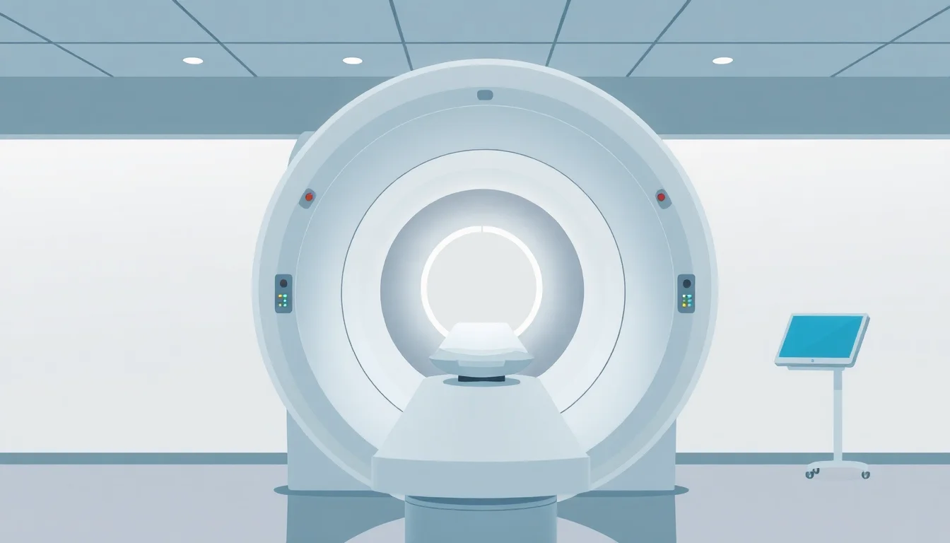
A typical MRI scanner comprises several critical components. The main structure is the magnet bore, a large, cylindrical tube housing a powerful superconducting superconducting magnet that generates the primary magnetic field, often ranging from 1.5 to 3 Tesla (T). Inside the bore, gradient coils create smaller, rapidly changing magnetic fields that allow for precise spatial localization of the signals. Radiofrequency coils, both built into the scanner and sometimes placed directly on the patient, transmit the RF pulses and receive the emitted signals. A sophisticated imaging workstation then processes this raw data, applying complex algorithms to reconstruct the images. The patient lies on a movable couch that slides into the bore, positioning the area of interest within the magnetic field's optimal range.
🏥 Who Needs an MRI and Why?
MRIs are indispensable for diagnosing a wide array of conditions. They excel at visualizing soft tissues like muscles, ligaments, tendons, cartilage, and nerves, making them crucial for orthopedic imaging and spinal diagnostics. In neurology, MRIs are vital for detecting tumors, strokes, multiple sclerosis, and other brain abnormalities. They are also used to examine organs such as the heart, liver, and kidneys, and play a significant role in cancer detection and staging. For pregnant women, the absence of ionizing radiation makes MRI a safer imaging option compared to X-rays or CT scans, particularly for fetal imaging when necessary. The detailed anatomical information provided aids clinicians in making precise diagnoses and tailoring treatment plans effectively.
⚡ MRI vs. Other Imaging: When to Choose What
When comparing MRI to other common imaging modalities, distinct advantages emerge. Computed Tomography offer faster imaging times and are excellent for visualizing bone and detecting acute bleeding, but they involve higher doses of radiation. Radiography are quick and cost-effective for bone fractures and certain lung conditions but provide limited detail for soft tissues. Ultrasound uses sound waves and is safe for pregnant women and for real-time imaging of organs and blood flow, but image quality can be operator-dependent and less detailed for deep structures. MRI's strength lies in its unparalleled soft tissue contrast and lack of ionizing radiation, making it the gold standard for many neurological, musculoskeletal, and oncological assessments, despite longer scan times and higher costs.
📈 The Evolution of MRI: From Giant Magnets to AI
The journey of MRI technology began with the foundational work on nuclear magnetic resonance in the mid-20th century. Early clinical MRI scanners, developed in the late 1970s and early 1980s by pioneers like Paul Lauterbur and Peter Mansfield, were large, cumbersome, and produced relatively low-resolution images. Over the decades, advancements in magnet technology, gradient coil design, and computer processing have dramatically improved image quality, speed, and patient comfort. The introduction of artificial intelligence is now revolutionizing MRI, enabling faster scan times through advanced reconstruction techniques, improved image analysis, and more accurate diagnoses. This continuous innovation promises to make MRI even more accessible and powerful.
⚠️ Safety First: What to Know Before Your Scan
Patient safety is paramount during an MRI scan. The powerful magnetic field means that any ferromagnetic objects (metals that can be magnetized) must be kept far away from the scanner. This includes pacemakers (unless MRI-compatible), certain implants, jewelry, and even some clothing with metallic threads. Patients are screened rigorously for any such contraindications. The loud, percussive noise produced by the gradient coils requires earplugs or headphones for comfort and to prevent hearing damage. Claustrophobia can be an issue in the enclosed bore, leading some patients to opt for open MRI scanners or to receive mild sedation. It's crucial to communicate any concerns or medical conditions to the technologist before the scan begins.
💰 Cost Considerations and Insurance
The cost of an MRI scan can vary significantly depending on the body part being imaged, the complexity of the scan, the geographic location, and the facility. Out-of-pocket expenses can range from several hundred to several thousand dollars. Medical insurance typically covers medically necessary MRIs, but coverage details, deductibles, and co-pays differ by plan. It's advisable to verify coverage with your insurance provider and the imaging center beforehand. Some centers offer discounted rates for patients paying out-of-pocket. Comparing prices between different facilities can lead to substantial savings, especially for elective or non-emergency scans.
🌟 Ratings and Reviews: What Patients Experience
Patient experiences with MRI scans are often characterized by the machine's noise and the confined space. Ratings frequently highlight the professionalism and attentiveness of the MRI technologists, who play a crucial role in patient comfort and ensuring the scan is performed correctly. While some patients report feeling anxious or claustrophobic, many find the experience manageable, especially with the use of headphones and clear communication from staff. The diagnostic value of the images produced is consistently rated highly, as MRIs often provide definitive answers that other imaging methods cannot. Online reviews and patient forums can offer insights into specific centers and technologists, helping prospective patients choose a facility that prioritizes comfort and quality care.
🚀 The Future of MRI: Faster, Smarter, Smaller?
The future of MRI technology is focused on enhancing speed, improving resolution, and expanding accessibility. Innovations like ultra-high field scanners (7T and above) are pushing the boundaries of anatomical detail, offering unprecedented insights into neurological disorders and research applications. Efforts are underway to develop more compact and potentially lower-cost MRI systems, including mobile units, to bring advanced imaging to underserved areas. The integration of quantitative imaging techniques and advanced computational analysis, including AI-driven image reconstruction and interpretation, promises to make scans faster, more precise, and more informative. The goal is to move beyond static images to dynamic, functional, and predictive diagnostics, transforming how we understand and treat disease.
Key Facts
- Year
- 1977
- Origin
- The first MRI scanner, known as the 'Indomitable,' was developed by Dr. Raymond Damadian and his team, with the first human scan performed in 1977.
- Category
- Medical Technology
- Type
- Technology
Frequently Asked Questions
Is an MRI scan painful?
An MRI scan is generally not painful. The primary discomforts reported by patients are the loud noise from the machine and the feeling of being in a confined space. Technologists can provide hearing protection and may offer mild sedation for those experiencing anxiety or claustrophobia. The magnetic field and radio waves used do not cause pain.
How long does an MRI scan typically take?
The duration of an MRI scan varies depending on the body part being examined and the specific protocols used. A typical scan can range from 15 minutes to over an hour. More complex scans, such as those involving the brain or spine, or requiring multiple sequences, will naturally take longer than scans of extremities like a knee or ankle.
Can I move during an MRI scan?
It is crucial to remain as still as possible during an MRI scan. Movement can blur the images and compromise their diagnostic quality, potentially requiring the scan to be repeated. Technologists will instruct patients on when it is safe to move slightly between image acquisitions, but during the actual scanning sequences, stillness is essential.
What are the risks associated with MRI scans?
MRI is considered a very safe imaging modality with no known long-term health risks from the magnetic fields or radio waves. The primary risks are related to ferromagnetic materials. Patients with certain metallic implants (e.g., older pacemakers, cochlear implants, aneurysm clips) may not be able to undergo an MRI due to the risk of the magnetic field moving or heating these objects. Screening is rigorous to prevent these issues.
What is the difference between an open MRI and a standard (closed) MRI?
Standard MRI scanners have a narrow, tube-like bore, which can be challenging for claustrophobic patients or larger individuals. Open MRI scanners have a more accessible design, with the magnet positioned above and below the patient, allowing for more open space. While open MRIs can improve patient comfort, they may sometimes produce lower-resolution images or take longer than closed systems, depending on the magnet strength and design.
Can I eat or drink before an MRI scan?
For most MRI scans, you can eat and drink normally before the procedure. However, for certain abdominal or pelvic MRIs, you may be asked to fast for a few hours beforehand or to drink a contrast agent. It's always best to confirm specific dietary instructions with the imaging center when scheduling your appointment.